
In today’s society, adult spinal deformity (ASD) is a well-known cause of intense pain and reduced mobility, often associated with the natural aging process and spinal column degeneration. This condition becomes increasingly prevalent with age, impacting nearly 68% of individuals over 60. In the broader context of healthcare, leveraging advanced technological tools like advanced computational imaging is necessary for managing the growing number of patients amidst the ‘Silver Tsunami.’
Advanced computational imaging techniques represent just one facet of how technology can be employed effectively. For example, AI-powered algorithms are demonstrating superior abilities in identifying specific cancers or pathologies compared to standard radiology techniques, highlighting technology’s potential to significantly enhance patient outcomes throughout a variety of medical conditions.
With the population above age 60 expected to nearly double by 2050, the prevalence of adult spinal deformity is becoming more pronounced, underscoring the urgent need for healthcare systems to integrate innovative technologies such as advanced computational imaging techniques to address this escalating issue.
Advanced Computational Visualization Techniques
The complexity of adult spinal deformity cases points to the importance of precision in appropriate patient selection and procedure execution. In recent years, research has highlighted the potential of artificial intelligence and machine learning in reshaping pre-operative preparations, surgical planning, and intra-operative navigation.
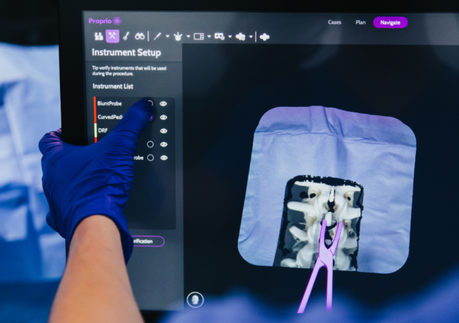
Enter the world of advanced visualization techniques, where light field technology is illuminating a path to a deeper understanding of patient anatomy, surgeon methodology, and surgical environments. Traditionally, spine surgery navigation has depended on surgeons combining pre-operative imaging with intra-operative anatomy to execute their treatment plans. However, with light field technology and depth sensors, these technologies digitize the entire operative field, merging it seamlessly with preoperative imaging, effectively bridging the gap between unseen and visible physical anatomy of the spine. This provides surgeons with a three-dimensional anatomical view that exceeds what the naked eye can discern.
Light field technology captures both the intensity and direction of light rays, crafting dynamic 3D representations of surgical scenes. This minimizes the need for repeated imaging radiation but also augments surgical precision, enhancing the boundaries between unseen and visible elements of surgery. The combination of these data sources creates a digital twin that unlocks novel insights and unlocks an entirely new approach to visualization. For instance, it allows surgeons to visualize three-dimensional structures beneath the skin or superimpose specific preoperative plans onto the digital scene, offering precise guidance during procedures. This meticulous level of detail empowers surgeons to make highly informed decisions, allowing complex procedures to be performed with heightened accuracy and ultimately, enhanced patient outcomes.
A Precise Approach to Intraoperative Alignment
Accurately assessing alignment during spine surgery is paramount to achieving preoperative goals. This allows surgeons to make adjustments, significantly reducing the risk of post-surgery complications or the necessity for revision surgery. While it’s widely acknowledged that achieving patient-specific intraoperative alignment yields optimal patient outcomes, current methods for assessing alignment during surgery are suboptimal due to challenges such as variability, time, and the requirement for ionizing radiation, resulting in limited utilization.
A recent study titled “Optical-kinematic measurement of spinal alignment: a radiation-free technique using light field navigation” showed an AI-powered algorithm in surgical measurement that outperformed surgeons, showcasing its efficacy and potential to revolutionize intraoperative procedures. The study recorded AI-measured and surgeon-measured vertebral coordinates in both pre-corrected and post-corrected states, capturing lateral fluoro shots before and after correction to provide surgeons with images to measure.
An AI-powered algorithm automatically calculated regional and segmental sagittal angles based on these coordinates. Seven spine surgeons evaluated 28 scans each, revealing a mean absolute difference of 1.96° between automated AI measures and manual measures. There was a strong correlation (r=0.98) observed between automated and manual measures, with 95% of all data falling within 2.53° of the mean manual measures.
Additionally, AI measurement significantly outpaced manual measurements performed by surgeons in terms of speed and eliminated the variability between the surgeon’s manual measures. These results highlight the accuracy and reliability of an AI-powered algorithm in achieving standard practice, pointing to its potential to revolutionize intraoperative spinal alignment assessments.
Integrating this technology into intraoperative alignment techniques is an advancement in precision and outcomes optimization for spine surgery. This provides accurate alignment measurements with minimal radiation exposure while streamlining workflow, reducing variability, and enhancing overall efficiency. Surgeons will always retain final clinical decision-making power regarding AI-powered guidance, ensuring that they receive the right information at the right time during procedures to make informed decisions and corrections.
Challenging Traditional Approaches: AI-Driven Decision-Making
For surgeons, the reliance on personal experience and shared knowledge has been a hallmark of spinal deformity surgery decision-making. However, the clinical results are highly variable and the complexities of each case necessitate a more data-driven and precise approach. This is where AI emerges as a powerful ally. Machine learning and deep learning models are capable of analyzing a diverse array of surgical cases and variables, providing standardized insights that inform treatment strategies. AI’s adaptability to unique patient profiles and learning from past cases empowers surgeons with data-driven decision support, ultimately improving both decision-making processes and patient outcomes.
Combining AI, Advanced Visualization, and Surgical Navigation
The integration of AI-supported algorithms and advanced computational imaging tools marks a significant advancement in patient care. These technologies enhance precision and have the potential to optimize outcomes while addressing the challenges posed by the growing prevalence of ASD among aging populations. By harnessing AI-powered tools combined with computational imaging, surgeons can achieve accurate alignment measurements with minimal radiation exposure, a streamlined workflow, reduced variability, and enhanced overall efficiency. This approach, grounded in a deep understanding of patient anatomy and surgical environments through light field technology, advances spinal deformity surgery by enhancing personalized care and improving patient outcomes.
Related Articles
-

Nearly 2/3 of device makers say their regulatory intelligence may be insufficient and they lack…
-

Cevian Capital increased its position in Smith & Nephew to a 5% stake in the British…
-

“In the future, CDRH intends to increase the breadth of the dataset of chemicals and…
-

With more than 50 years in the medical device space, SERF SAS is recognized for…