Three Areas Where Tissue Engineering Is Changing Medical Treatment

Tissue engineering encompasses the fields of engineering materials, chemical factors and living cells. Recent breakthroughs in 3-D printing technology allow finer tuning of parameters when designing tissues and scaffolds. Not only are base materials being predictably constructed, but bioengineers can now seed and build-in living cells.
3-D printing processes enable the introduction of drugs into materials, allowing them to act as a timed-release medium seeded by, or implanted into, living cells and tissue. Research has exploded over the past decade, moving quickly towards real-life applications—from edible lab burgers to new skin for burn victims. Tissue engineers are also working towards replacing damaged or diseased natural organs with lab-grown organs.
Tissue Scaffolding
The first step of creating functional tissue is to provide a viable matrix or scaffold to accommodate their growth and integration. A range of established methods to build 3-D biomaterial scaffolds exists—from hydrogels to emulsification freeze-drying and laser-assisted bioprinting. Electrospinning arguably provides the best platform for control of scaffold parameters and cellular growth. This is due to the ability to tune the fibers’ diameter, location, orientation, number-density, drug-load, inherent material properties (e.g. porosity), process cleanliness, scaffold built dimensions and overall 3-D shape. Electrospinning is considered a type of 3-D printing; similar to some other 3-D printing processes, all electrospinning pushes a liquid through a nozzle to create filament that is pulled onto a grounded platform (drum or plate).
The three main types of electrospinning are: Coaxial, Emulsion and Melt. Each has its merits and drawbacks. Coaxial makes use of two materials, one as a core extrusion and the other as an annular sheath over the core. The core can be either a polymer, a drug, or a combination, depending on the application. The external sheath is usually a structural, biodegradable polymer. Emulsion electrospinning makes use of a polymer that is dispersed in a solvent, with or without drugs, and is separated from the solvent through evaporation.
Once a viable scaffold is fabricated, cells can be integrated into it. This can be done through manual seeding into a solution or via bio-ink based printing. Examples of tissues grown with scaffolds cover all of the body’s organs, including in-vitro livers, bladders, kidneys, hearts and lungs. General tissues such as nerves, cartilage, skin, bone, blood vessels and muscle can also be printed. Over time, most scaffolds will biodegrade and be replaced by cells’ extracellular matrix. This makes them great candidates for implants as no residual material remains in the long term.
Nerve Grafting
Early-stage research using microfiber scaffolds has shown promise in cultivating and directing the growth of nerve cells—motor neurons in particular. Providing known topographies, pathways of growth can be fabricated into the scaffold, and seeded cells will produce dendrites that “crawl” along these fibers. These dendrites seek other cells’ dendrites, which they then fuse with, eventually resulting in a network of functional motor neurons. The intended goal of the research is to spark a pathway toward viable nerve grafts for breaks in the central nervous system (glial scars), which could result in solutions to disabilities such as lower body paralysis.
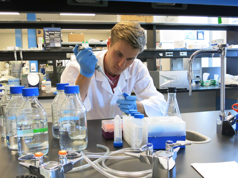
I had the pleasure of being involved in several experiments that looked into this research within the Willerth Lab at the University of Victoria. These experiments culminated in a published paper that demonstrated the ability of both micro and nanofibers’ abilities to act as effective scaffolds for neuronal differentiation and organized propagation. Both melt and solution electrospinning were employed, with the latter including retinoic acid (a differentiation promotion factor) that was incorporated and provided a timed release. The findings provided a sound basis for further research into how physical and chemical cues can be used to engineer neural tissue.
Skin Printing
Breakthrough work by several labs is bringing a novel conept to life—the real-time printing of skin directly onto patients. This could have wide implications for the medical field, from burn victim skin repair to plastic surgery. Because printers are portable, potentially low maintenance, and clean, they could soon find their way into surgical rooms. The development of consistent printed skin has been progressed by labs such as that of Dr. Marc Jeschke, who amongst other titles is a Director of the Ross Tilley Burn Centre at Sunnybrook Health Sciences. He describes current skin grafting techniques as “barbaric”, with the need to peel skin from elsewhere on a patient’s body to replace that lost in burned regions. This skin usually comes from the legs or back, and is extracted painfully using a dermatome, or skin planer. The healing process following surgery for both burn and graft extraction area is painful and takes months before fully healing. It is also prone to infection, as the area stays moist and exposed during healing.
Within Dr. Jeschke’s lab, skin printing takes place on their prototype, the “PrintAlive” Bioprinter. It makes use of an extruded gel containing cells within it, which also serves as the extracellular matrix during growth. This is printed onto a drum, which pulls and elongates the skin analogue to make a resultant strip. The strip is then collected and cultured. The future goal is to refine and use the process on a larger scale with patient-derived cells to tailor grafts on an as needed basis.
Consensus among bioengineers is that skin is the easiest organ to print; however, the elusive challenge remains of how to vascularize the tissue. By nature, vasculature is well-integrated into tissue by gradually growing with it—whether from scratch in the womb or by nurture such as through muscle growth. With printing, this is more difficult, as tissue is built rapidly and completely, with the expectation that it becomes fully functional within hours. Vasculature requires a full fluid pathway to be effective, as recirculation is needed to remove waste and add nutrients. This remains the biggest nut to crack before skin (or any) tissue engineering takes off.
Conclusion
While tissue engineering is driving fast towards real-life applications, the research and development of viable tissues and treatments is still progressing. Once ready, clinical trials demonstrating the in situ treatments with bioengineered tissue will be instrumental to their introduction into medicine.